Cleanly exfoliated 2D materials are the foundation of high-quality van der Waals heterostructures. There are plenty of tricks that can improve the process, but ultimately, nothing replaces experience. Of course, using the right exfoliation tape and a clean substrate also goes a long way toward achieving reliable, high-quality flakes.
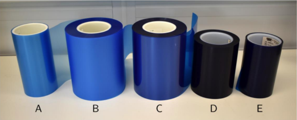
Choose your tape: This has been a long-standing debate in the 2D materials community, with some research groups using blue cleanroom tape and others relying on regular Scotch tape. In practice, there is no clear consensus that one is universally better than the other, as both approaches can produce clean, high-quality flakes when properly handled.
As with many aspects of exfoliation, experience plays a key role. It is worth testing different tapes and developing your own process, as the results can depend on several factors, including the humidity and temperature of the room where the exfoliation is performed.
Heating technique: To obtain large-area monolayer and bilayer graphene, as well as other high-quality 2D materials, it is highly recommended to use the approach developed in this article. In brief, the Si/SiO₂ substrates should first be cleaned using a gentle oxygen plasma (~20 s) treatment before bringing the exfoliation tape into contact with the substrate. Once the tape carrying the exfoliated material is placed in contact with the substrate, the sample is heated on a hot plate at approximately 105 °C for ~4 minutes. After heating, allow the sample to cool down before carefully peeling off the tape, leaving the exfoliated flakes on the substrate.
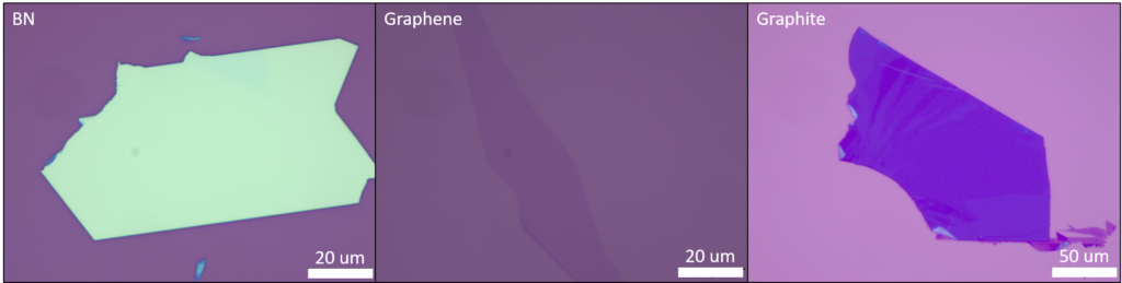
BN exfoliation: when relatively thick flakes of BN (30 nm – 80 nm) are desired, the process is similar to the one described above, but without heating the substrate before peeling off the tape. Avoiding the heating step generally results in cleaner BN flakes improving the quality of the exfoliated material.
Pro tips:
- If using a reactive ion etcher (RIE) for plasma cleaning, make sure that the plasma treatment is not too aggressive. Excessive plasma power or long exposure times can make the subsequent pickup process much more difficult. In our experience, an oxygen plasma treatment of 20–30 seconds is usually sufficient.
- While the chips are on the hot plate, gently press the tape with your thumb to improve the contact between the exfoliated material and the substrate. Better contact generally leads to improved flake transfer.
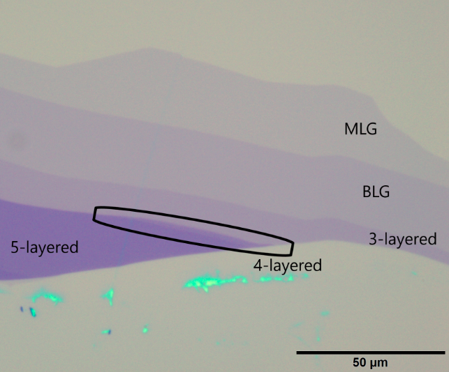
Identifying the number of layers: After exfoliation, the number of layers in a flake can be estimated from its optical contrast when observed under an optical microscope. The contrast depends on several parameters, including the 2D material itself (graphene, BN, etc.) and the substrate properties (for example, the thickness of the SiO₂ layer). Careful calibration and comparison with known layer numbers are often required for accurate identification [reference article]. When high enough, the contrast will present discrete steps whose amplitude is directly linked to the number of layers.
Pro tips:
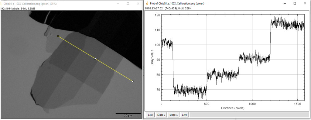
The optical contrast of a monolayer can be quantified using image analysis software such as ImageJ. To do this, first separate the image into its individual color channels (red, green, and blue), then draw a line profile across regions with different flake thicknesses, using the green color. By analyzing the intensity variation along this profile, the contrast associated with the monolayer can be extracted and compared with thicker regions to identify the number of layers.
Finally, Raman spectroscopy can be particularly useful to identify monolayers [reference article].
Lastly in some cases, given any combination of substrate and light wavelength, the optical contrast can remain low, this is for example the case of monolayer BN, and other methods are necessary to identify monolayers.
Particularly challenging: monolayer BN
Monolayer and few-layer BN are especially difficult to obtain and identify using optical microscopy due to their extremely low optical contrast on standard substrates. In our experience, and as previously reported, the most reliable approach to obtain monolayer BN is to use the heating-assisted exfoliation technique described above.
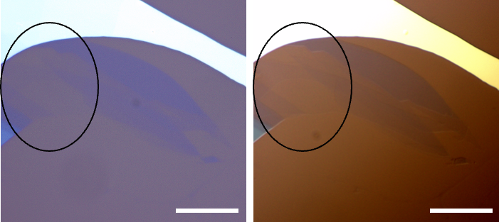
Performing the exfoliation on a 90 nm SiO₂ substrate provides sufficient optical contrast to identify monolayer flakes. Their visibility can be further enhanced by using a U-DICR (ultra-differential interference contrast) polariser, which improves the contrast between the thin BN flakes and the substrate.
TMDs Exfoliation
Under construction
Au assisted Exfoliation
Under construction
Cryogenic exfoliation (rhombohedral graphene)
Under Construction
Air sensitive material exfoliation (gloves box environment)
Under Construction